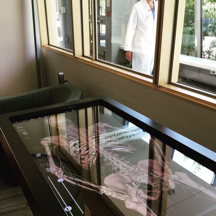
This Table is an advanced visualization system for anatomy education that uses touch screen capabilities and offers virtual dissection. Designed to fit a life-sized image, the human body can be manipulated to show different anatomical sections with the ability to rotate and view the body or part from all angles. Layers of the body can be removed, certain sections isolated, and cross-sections made, all with pinning, labeling, and color-coding capabilities, among many other functions.
Professors and students have the ability to save a manipulated image for teaching purposes, examinations, or presentations. The Table provides curriculum-based tools, import/export capabilities, and an extensive archive of virtual images: full body and regional, CT scans, Histology, and case studies. The Table also has projection capabilities when hooked up to a separate computer and screen system.
The Table will be on display in the front of the library through the end of September. For training on the basic of how to use the Table, please contact Kate Bright to set up a session.
